
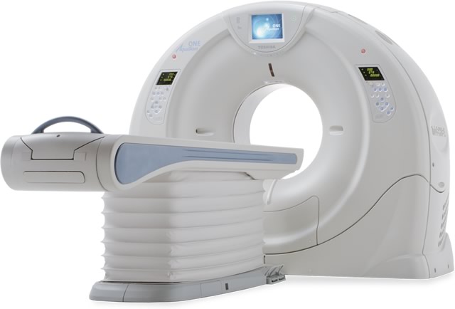
We use the best-in-class Toshiba Aquilion ONE 320 slice computed tomography (CT) scanner. The scanner is very fast and can perform advanced cardiac imaging and neuroimaging.
We emphasize radiation safety with tailored exams and the use lowest possible radiation doses.
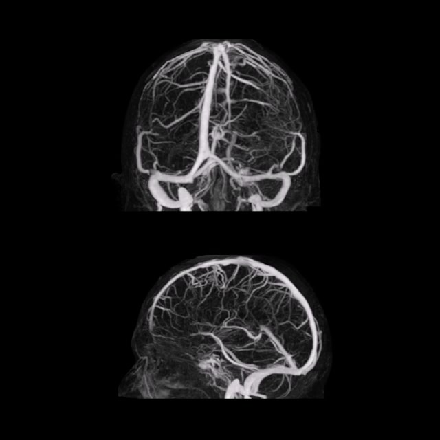
Neuro CT
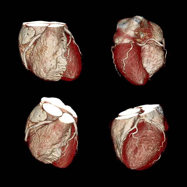
Cardiac CT
The term “computed tomography”, or CT, refers to a computerized x-ray imaging procedure in which a narrow beam of x-rays is aimed at a patient and quickly rotated around the body, producing signals that are processed by the machine’s computer to generate cross-sectional images—or “slices”—of the body. These slices are called tomographic images and contain more detailed information than conventional x-rays. Once a number of successive slices are collected by the machine’s computer, they can be digitally “stacked” together to form a three-dimensional image of the patient that allows for easier identification and location of basic structures as well as possible tumors or abnormalities.
Unlike a conventional x-ray—which uses a fixed x-ray tube—a CT scanner uses a motorized x-ray source that rotates around the circular opening of a donut-shaped structure called a gantry. During a CT scan, the patient lies on a bed that slowly moves through the gantry while the x-ray tube rotates around the patient, shooting narrow beams of x-rays through the body. Instead of film, CT scanners use special digital x-ray detectors, which are located directly opposite the x-ray source. As the x-rays leave the patient, they are picked up by the detectors and transmitted to a computer.
Each time the x-ray source completes one full rotation, the CT computer uses sophisticated mathematical techniques to construct a 2D image slice of the patient. The thickness of the tissue represented in each image slice can vary depending on the CT machine used, but usually ranges from 1-10 millimeters. When a full slice is completed, the image is stored and the motorized bed is moved forward incrementally into the gantry. The x-ray scanning process is then repeated to produce another image slice. This process continues until the desired number of slices is collected.
Image slices can either be displayed individually or stacked together by the computer to generate a 3D image of the patient that shows the skeleton, organs, and tissues as well as any abnormalities the physician is trying to identify. This method has many advantages including the ability to rotate the 3D image in space or to view slices in succession, making it easier to find the exact place where a problem may be located.
CT scans can be used to identify disease or injury within various regions of the body. For example, CT has become a useful screening tool for detecting possible tumors or lesions within the abdomen. A CT scan of the heart may be ordered when various types of heart disease or abnormalities are suspected. CT can also be used to image the head in order to locate injuries, tumors, clots leading to stroke, hemorrhage, and other conditions. It can image the lungs in order to reveal the presence of tumors, pulmonary embolisms (blood clots), excess fluid, and other conditions such as emphysema or pneumonia. A CT scan is particularly useful when imaging bone fractures, joints, cartilage, or tendons since it usually produces more detail than would be possible with a conventional x-ray.
As with all x-rays, dense structures within the body—such as bone—are easily imaged, whereas soft tissues vary in their ability to stop x-rays and, thus, may be faint or difficult to visualize. For this reason, contrast agents have been developed that are highly visible in an x-ray or CT scan and are safe to use in patients. Contrast agents contain substances that are better at stopping x-rays and, thus, are more visible on an x-ray image. For example, to examine the circulatory system, a contrast agent based on iodine is injected into the bloodstream to help illuminate blood vessels. This type of test is used to look for possible obstructions in blood vessels, including those in the heart. Other contrast agents, such as barium-based compounds, are used for imaging the digestive system, including the esophagus, stomach, and GI tract.
All x-rays produce ionizing radiation, which has the potential to cause biological effects in the human body. For patients, these biological effects can range from an increased lifetime risk of cancer to possible allergic reactions or kidney failure due to contrast agents. Under some rare circumstances of prolonged, high-dose exposure, x-rays can cause adverse health effects such as skin reddening (erythema), skin tissue injury, hair loss, cataracts, or birth defects (if scanning conducted during pregnancy).
For conventional x-rays, the amount of radiation delivered to a patient is extremely small. However, for a CT exam, such as a study of the abdomen, the radiation delivered to the patient can be equivalent to as many as 400 chest x-rays. Similarly, a CT exam of the head can produce the equivalent of about 100 chest x-rays. For this reason, it’s important that CT exams are limited only to those cases where the benefit to be gained greatly outweighs the increased risk. This is especially true for children, who are more sensitive to ionizing radiation and have a longer life expectancy and, thus, have a higher relative risk for developing cancer than adults. Also, a child’s smaller size affects the amount of radiation dose received. For this reason, when scanning children, equipment settings need to be adjusted to reduce the radiation dose while maintaining high image quality.
Provided by the National Institute of Biomedical Imaging and Bioengineering
For additional information, visit radiologyinfo.org - Computed Tomography