
We use the best-in-class Siemens 3T MAGNETOM Skyra scanner with new software that enables rapid imaging, such as completing a brain scan in 5 five minutes or less. This scanner also has advanced neuroimaging, musculoskeletal, breast, and cardiac imaging capabilities.
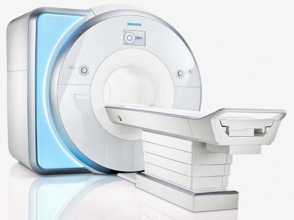
MAGNETOM Skyra
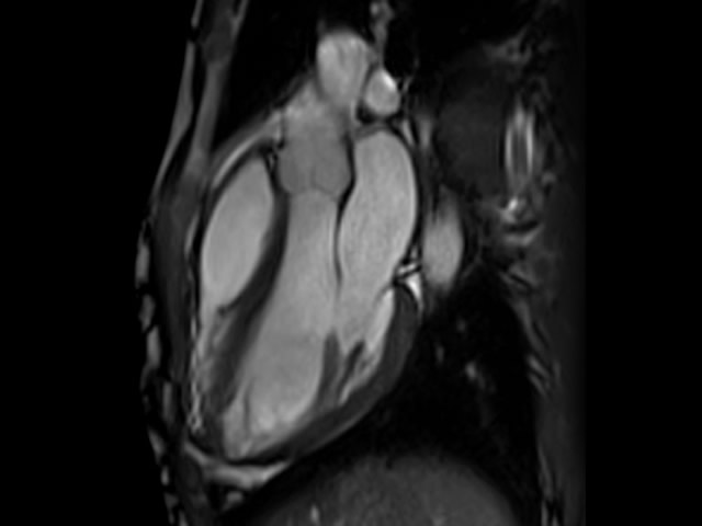
Cardiac MRI
Advanced noise reduction technologies are present on all MAGNETOM® scanners. Nevertheless, we have continuously strived to develop technologies that will further lower noise without compromising imaging efficiency and quality. With Quiet Suite, we have addressed the root source – sharp gradient switches – to take noise reduction to a new level. Quiet Suite includes QuietX sequences and the inaudible PETRA as well as optimized protocols for neurological and orthopedic examinations. Our latest innovation – QuietX Diffusion1 – adds diffusion imaging to our quiet portfolio. Experience a broad range of benefits:
GOBrain delivers reliable quality at exceptional speed. It enables clinically validated, push-button brain exams, with multiple orientations and all relevant contrasts – in only five minutes1. This fast exam is more tolerable for patients, and helps potentially reduce motion-related artifacts and the need for rescans and sedation. As a result, GOBrain could double throughput and reduces costs per scan
Magnetic resonance imaging, or MRI, is a non-invasive imaging technology that produces three dimensional detailed anatomical images without the use of radiation. It is often used for disease detection, diagnosis, and treatment monitoring. It is based on sophisticated technology that excites and detects the change in the direction of the rotational axis of protons found in the water that makes up living tissues.
MRIs employ powerful magnets which produce a strong magnetic field that forces protons in the body to align with that field. When a radiofrequency current is then pulsed through the patient, the protons are stimulated, and spin out of equilibrium, straining against the pull of the magnetic field. When the radiofrequency field is turned off, the MRI sensors are able to detect the energy released as the protons realign with the magnetic field. The time it takes for the protons to realign with the magnetic field, as well as the amount of energy released, changes depending on the environment and the chemical nature of the molecules. Physicians are able to tell the difference between various types of tissues based on these magnetic properties.
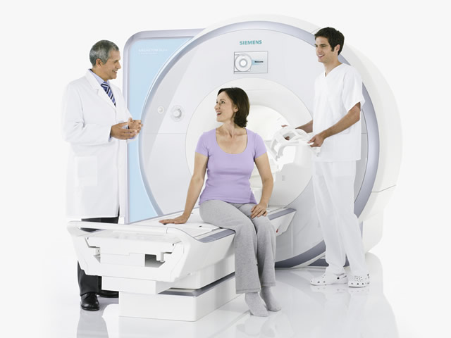
To obtain an MRI image, a patient is placed inside a large magnet and must remain very still during the imaging process in order not to blur the image. Contrast agents (often containing the element Gadolinium) may be given to a patient intravenously before or during the MRI to increase the speed at which protons realign with the magnetic field. The faster the protons realign, the brighter the image.
MRI scanners are particularly well suited to image the non-bony parts or soft tissues of the body. They differ from computed tomography (CT), in that they do not use the damaging ionizing radiation of x-rays. The brain, spinal cord and nerves, as well as muscles, ligaments, and tendons are seen much more clearly with MRI than with regular x-rays and CT; for this reason MRI is often used to image knee and shoulder injuries.
In the brain, MRI can differentiate between white matter and grey matter and can also be used to diagnose aneurysms and tumors. Because MRI does not use x-rays or other radiation, it is the imaging modality of choice when frequent imaging is required for diagnosis or therapy, especially in the brain. However, MRI is more expensive than x-ray imaging or CT scanning.
One kind of specialized MRI is functional Magnetic Resonance Imaging (fMRI.) This is used to observe brain structures and determine which areas of the brain “activate” (consume more oxygen) during various cognitive tasks. It is used to advance the understanding of brain organization and offers a potential new standard for assessing neurological status and neurosurgical risk.
Although MRI does not emit the damaging ionizing radiation that is found in x-ray and CT imaging, it does employ a strong magnetic field. The magnetic field extends beyond the machine and exerts very powerful forces on objects of iron, some steels, and other magnetizable objects; it is strong enough to fling a wheelchair across the room. Patients should notify their physicians of any form of medical or implant prior to an MR scan.
When having an MRI scan, the following should be taken into consideration
Provided by the National Institute of Biomedical Imaging and Bioengineering
For additional information, visit radiologyinfo.org - Magnetic Resonance Imaging (MRI)